About Cracked Tooth Syndrome
- Causes
- Signs and Symptoms(features)
- Treatment
As you can see the symptoms above vary and are not always the same for everyone. On top of this, the pain history can be similar to other conditions as a result the dentist may need to do the following to determine the cause.
Signs of wear be that crazed lines, worn down cusps. The bite may have missing teeth which is placing more force on others. Are there large fillings that may have weakened cusps
Due to the difficulty you may at pinpointing the pain. The dentist will want to apply some tests to replicate the pain in order to locate the tooth causing the issue. Bite tests are very useful in determine the tooth in question. Due to the difficulty often in the patient localising where the pain is coming from, as a result this device is designed to localise pressure to individual cusps so that when you bite down you cause flexion on the crack which is thought to allow fluid to move in and stimulate your sharp nerve fibres. Therefore if there is a crack your likely to feel it when you bite down or release biting pressure on this instrument. The dentist may also use ice or hot/cold water to replicate the pain also if mentioned in your pain history.
As you can see the symptoms above vary and are not always the same for everyone. As mentioned above the pain history can be similar to other conditions as a result the dentist may need to do the following to determine the cause.
If the tooth is a house/car the gums are like the foundations/wheels, they tell a lot about the options we have long term for the tooth. Isolated deep probing depths may be signs that the crack has gone below the gums. These results do play a big role in determining the long term options for the tooth.
Sometimes the dentist may apply a coloured dye in order to better visualise any cracks on the suface of a suspect tooth
The light will move through if there are no cracks. However, if a crack is present it will block the light.
Just like a house, you sometimes need to remove some of the carpet to see the hard wood floors underneath. The same is with cracked teeth. If the suspect tooth has a filling, the dentist may want to remove the old filling to be able to determine if a crack is present and to what extent and direction. As these do play a role in determining the long term options for the tooth.
This is used if the dentist has a provisional diagnosis of a cracked teeth. What I mean by this, is it is used to confirm the diagnosis of the painful symptoms you are having. How it works is that the metal band is like a band around a barrel, it hugs the tooth, by doing this you prevent the flexing of the cusp. Without the flexing we are not geting the fluid movement to the nerve and thus not getting the stimulated pain. (see the barrel picture above for a visual explanation) There is the downside of an unaesthetic appearance especially if the metal band is near the front of your mouth, it can affect the gums if not cleaned properly, as well as possibily of requiring the removal of tooth structure. Understand that the metal band is not a permanent solution and is why it needs to be reviewed in a few weeks to months time to decided if the tooth needs a permanent solution or further investigations are needed.
Uses the same concept of hugging/capping idea as the orthodontic band, but are used more often in teeth that have an old filling. Understand that these also need to be reviewed in a few weeks to months time to decided if the tooth needs a permanent solution or further investigations are needed.
There are many ways to classify cracked teeth, but here to make it easier to understand we will consider simple cracks and complex cracks. But treatment depends on multiple factors and is unique to every situation. It inolves detailed treatment planning and patients should be aware that even with the highest standards of dentistry, it may not be possible to save the cracked tooth.
Most often for these cases it involves removing weakened cusps and placing a large filling. If more than one cusp is fractured or if the tooth is heavily restored, often a crown is a more effective treatment option. As the crown protects the tooth and often prevents the crack from progressing.
Usually at this stage the crack has progressed into the pulp or caused irreversible damage by inflammation to the pulp. In these cases root canal treatment is usually required before needing to place a crown or filling. Follow the link for explaination of Root Canal Treatment. In some cases the case may be to complicated and requires specialist training and equipment. If this is the case the dentist may refer you to an endodontist (pulp (inside the tooth) specialist) or prosthodontist (crown (the top part of the tooth) specialist)
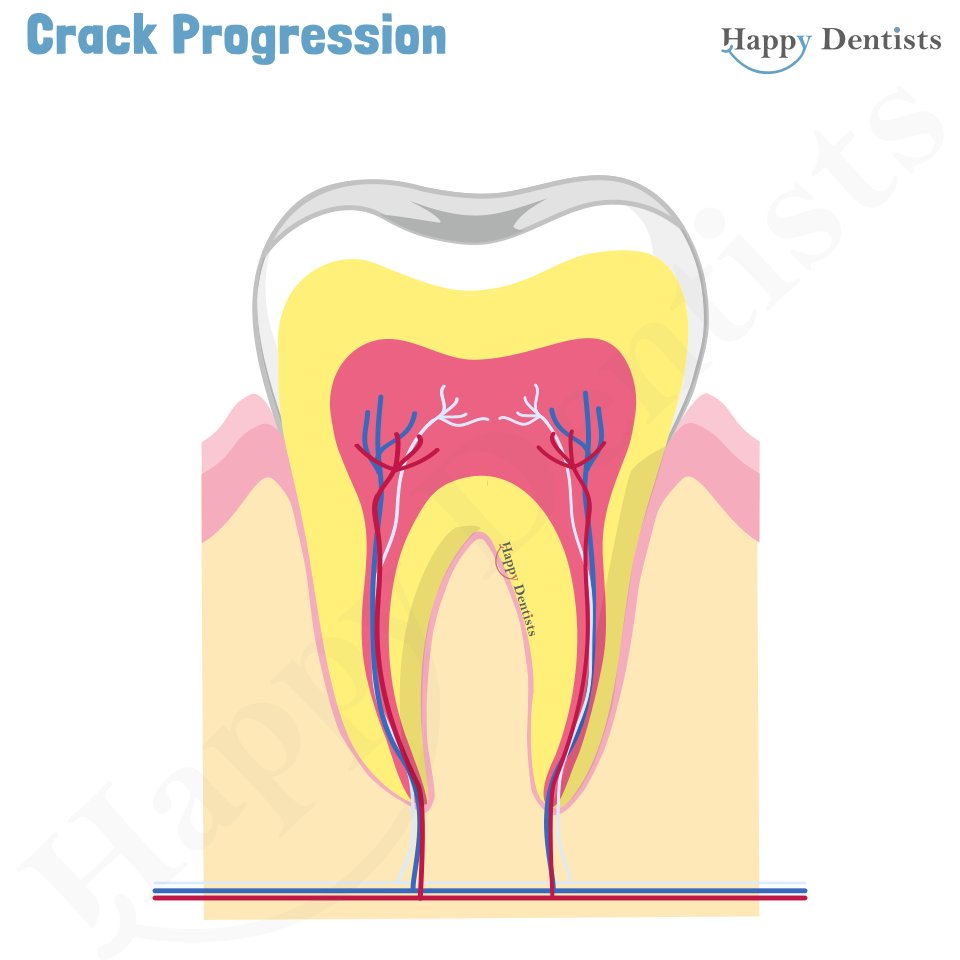
The longer a simple crack is left untreated (includes prevention treatment), there is a high liklihood that it can progress into a complex crack or fracture (like the picture above with the car). Everytime it progresses to the next stage it becomes hard and more complex to treat. Involving more time and cost. If gets to the pulp then root canal treatment will be necessary, or in some cases removal of the tooth. In severe cases, where the tooth has split in half, the tooth usually has to be removed. Then you may have to look at replacement options like a bridge, denture or dental implant.

If you have finished reading all the information on this page, get a certificate for your hard work.
This page provides general information about dental topics. It does not contain all the known facts of this subject and is not intended to replace personal advice from your dentist. If your not sure about anything on this site, contact us or speak to your local oral health practitioner. Make sure you give your local oral health practitioner your complete medical history and dental history.
A selection of the references used:
Hasan, S., Singh, K., and Salati, N. (2015). Cracked tooth
syndrome: Overview of literature. International journal of applied
and basic medical research, 5(3), 164–168.
https://doi.org/10.4103/2229-516X.165376
Kahler, W. (2008). The cracked tooth conundrum: terminology,
classification, diagnosis, and management. American journal of
dentistry, 21(5), 275.